Last Updated on: 19th December 2025, 09:13 am
Dental diseases in dogs
Diseases affecting dental growth and development in canines
By virtue of the dog’s status as a companion animal, there are many veterinary publications and reference texts on the diagnosis, medical management, pathology, and epidemiology of canine disorders.
Tooth disorders due to genetic or environmental reasons

It is well established that systemic diseases in dogs, particularly those associated with severe pyrexia, infection with epitheliotropic viruses, exposure to certain medications (such as tetracycline or high levels of systemic fluoride) during tooth development, can result in enamel abnormalities (1,2).
These abnormalities are responsible for the formation of a unique extracellular matrix, the maturation of the matrix, and its mineralization to form enamel. An optimal extracellular environment is required for this to happen normally (3).
Enamel hypoplasi and hypomineralization

Enamel hypoplasia is a generic term defined as quantitative enamel defects that present as foci of reduced enamel thickness.
1) Clinical findings
- Pitts
- Lines / grooves
- Areas or absence of enamel on the crown of a tooth
2) Enamel hypoplasia due to pathogenesis
- Hypoplastic enamel matrix production
- Matrix hypomineralization
3) Hereditary factors
- Amelogenesis imperfecta
4) Environmental factors
- Trauma
- Infectious
- Pyrexia of any origin
- Chemicals with toxic influence on ameloblasts (ie fluoride)
The clinical implications of enamel lesions as a result of any form of Enamel Hypoplasia (EH) include the potential for tooth sensitivity and an increased susceptibility to wear, erosion, and even cavities due to structurally defective enamel and the resulting plaque-retaining nature of the tooth surface.
Focal enamel or turner’s tooth hypoplasia
The management of these defects ranges from improved tooth brushing techniques and preventing plaque build-up on the rough surface of the defect to restoration of the defect. However, in cases where the defect extends below the gingival margin, gingivitis is common around the affected teeth. If left untreated, this can increase the risk of periodontitis. In cases where subgingival involvement prevents optimal restoration of the defect and where bone loss is already present, periodontal surgery is warranted prior to restoration or extraction.
Diffuse enamel hypoplasia
This dental condition is similar to the previous one; the difference is that in the case soutlined here, the presentation is generalized. It affects the teeth as a whole, and treatment requires comprehensive management.
Amelogenesis imperfecta
There is a wide variety of clinical presentations of AI with respect to the quantitative and qualitative aspect of the enamel.
Only two studies with confirmed cases of AI in dogs are available in the English literature. One group describes a familial type HE in standard poodles in Sweden that resembles a hypocalcified type AI clinically and histologically (4) but without genetic confirmation. The study included a four-generation pedigree, and a research project was carried out to determine the incidence of familial tooth discolorations in Swedish poodles.

Over two years, 16.1% of 62 Swedish Poodles were confirmed to have similar dental changes (4). Another group reported a familial enamel hypoplasia that uniformly affects deciduous and permanent teeth in Italian greyhounds. In humans, the clinical manifestations of AI often depend upon the type present. In the genetically confirmed case of IA in greyhounds, the enamel was thin, rough with brown spots, and the teeth were small and pointed with larger-than-normal interdental spaces (5). It was not possible to confirm the diagnosis of AI in any of these cases. Depending upon the extent of hypoplasia caused by AI, treatment will vary. However, the most important thing is genetic counseling with a recommendation to neuter the genetically-affected dog.
Tooth discoloration during development
Extrinsic stains in tooth enamel result from various types of pigments deposited on the enamel surface. They usually can be easily removed with inlaying and polishing procedures. Although a description of extrinsic tooth discoloration is beyond the scope of this article, it is important to know that extrinsic stains can be incorporated into the substance of the tooth, an entity known as internalized discoloration.
The intrinsic color of a tooth is influenced by the thickness and structural properties of the enamel that influence the scattering and absorption of light within the enamel. A variety of local and systemic causes can result in intrinsic tooth discoloration. Systemic causes include drug-related tooth discoloration, particularly tetracycline staining (see below), metabolic conditions such as fluorosis, and genetic conditions that can lead to tooth discoloration. These include congenital erythropoietic porphyria with accumulation of porphyrins and hyperbilirubinemia, which causes the deposition of by-products of hemolysis, as well as structural defects such as IA and dentinogenesis imperfecta (6).

Dentinogenesis imperfecta, on the other hand, represents a variety of rare genetic disorders that result in a variety of genetically abnormal dentin structures (6). Abnormal dentin thereby provides inadequate support with resulting enamel fractures and the exposure of porous dentin, prone to chromogen uptake. Dentin has an abnormal color, resulting in opalescent teeth.The local causes of intrinsic tooth discoloration are numerous and include pulp necrosis, pulp hemorrhage (within the pulp cavity), and pulp tissue debris after endodontic therapy, resulting in the deposition of hemoglobin-related pigments in dentin. Endodontic materials within the pulp chamber, as well as aging with secondary and tertiary dentin deposition on the pulp walls, also cause substantial discoloration of the teeth (6).
Tetrcycline staining
Tetracycline has been on the market for more than six decades and is commonly used to treat a variety of bacterial, chlamydial, mycoplasma, and rickettsial infections. One of the most adverse drug reactions from the use of tetracycline is the permanent discoloration of the teeth from yellow to brown, which affects the parts in the process of mineralization and matrix maturation at the time of use (7 )
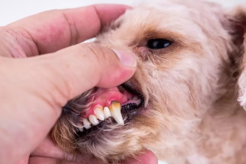
Periodontal disease
Periodontal disease (PD) is defined as an inflammatory response to dental plaque; it is the most common oral pathology in adult dogs and cats (8). The term, periodontium, refers to the tissues that surround and support the tooth. It is composed of the gum, cementum, periodontal ligament and alveolar bone. The main function is to join the teeth to the bone tissue of the jaws.
Several studies have reported a high frequency of PD in dogs, which have allowed us to know the role of dental plaque and other associated factors, such as age, size of the animal, head biotype, diet and chewing behavior. PD affects systemic health, as there is a positive correlation between the severity of PD and microscopic changes in the structural tissues observed in the heart, lungs, kidney and liver of dogs (9).
The frequency of periodontal disease (76.9%, 40/52) in the present study was similar to the results found in other countries (10). However, there is evidence of higher frequencies, such as the 97% reported by Gad, 1968 and the 90% found by Hoffmann and Gaengler (12) in dogs of the poodle breed, possibly due to the small size of the skull.
The teeth most frequently affected were the premolars, a finding that corroborates similar studies. When the dog does not receive frequent dental hygiene, the teeth, especially the premolars, accumulate plaque and dental stones due to the mouth of the canals of the salivary glands between the fourth premolar and first upper molar, favoring a constant mineral deposition for the rapid formation of dental tartar (9).
Self-cleaning due to the type of occlusion, the mechanical force of chewing, salivary flow, and tongue movement are inefficient in removing plaque and dental tartar in this type of tooth (11). In breeds with skulls, where the transverse diameter is greater than the longitudinal diameter, a rotation of the premolars is observed, allowing for the creation of a greater number of potential retentive surfaces of dental plaque and tartar (9)
Kyllar and Witter (11) point out that the upper teeth are more affected, while other researchers (10,12) found no difference between the upper and lower teeth. In the present study, the upper teeth were the most affected.
Contact Us for dental diseases in dogs
If you have any questions about this or other topics, contact us at Channel Island Family Dental and on our Facebook page. At Channel Island Family Dental, we are always attentive to your needs to make a timely diagnosis. In addition, our dentists in Oxnard, Santa Paula, Ventura, Newbury Park, and Port Hueneme will guide you to the best treatment to give you back your best smile.