Last Updated on: 5th January 2026, 06:12 am
In some people, abnormal cell growth may be observed in the oral cavity; it can be caused by various conditions like erythroplakia. This abnormal growth, known as proliferation or sometimes warts, can originate in any tissue inside or outside of the mouth.
Proliferations generally form on the lips, the sides of the tongue, the floor of the mouth, and the soft palate. Sometimes, they cause pain or irritation. Oral proliferations can be:
- Benign
- Premalignant (dysplastic)
- Malignant.
In this document, we will delve a little deeper into premalignant lesions, including how to identify them and what treatments exist.
What is a Premalignant Lesion?
Precancerous lesions are growths of cells that, although initially harmless, can become cancerous over time. In the mucosa of the mouth, three types of premalignant lesions can be distinguished:
1. Leukoplakia
This can appear as a white or gray patch or area that cannot be removed by scraping. It usually occurs when the oral mucosa is irritated for a prolonged period. The white area may appear thicker, as it has a thick layer of keratin. Most of the time, leukoplakia does not become cancerous, but it has been shown that 20% of leukoplakias when examined under a microscope, are cancerous or have cell changes (dysplasia) that can evolve into cancer if not properly treated.
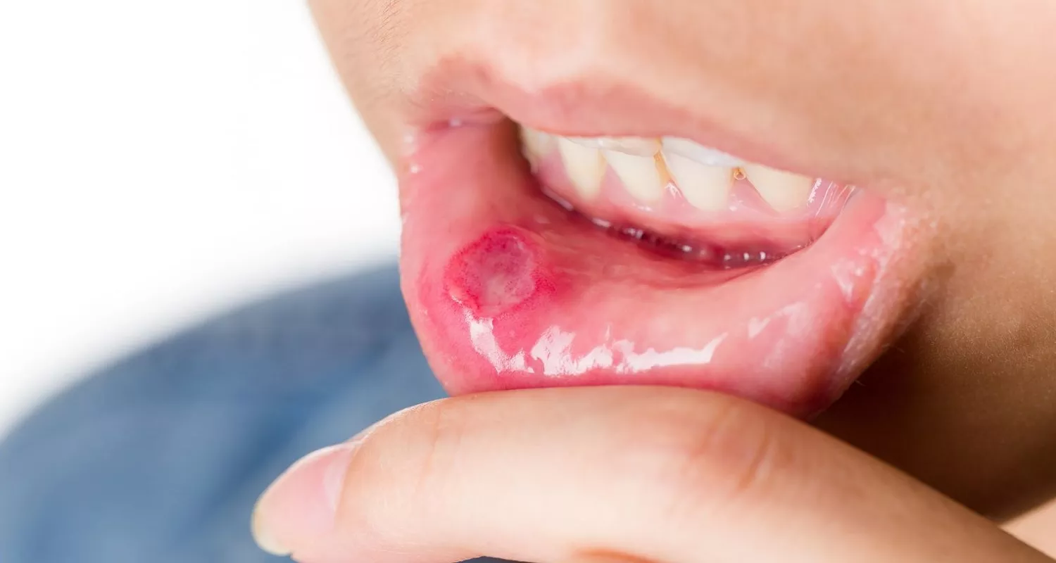
2. Erythroplakia
It is an abnormal area of flat, red tissue with a worn appearance that usually occurs when the oral mucosa wears away. It may or may not be slightly raised and may bleed when scraped. It has a velvety appearance and can manifest small white spots. It is less common than leukoplakia, but it is often more serious. In the vast majority of cases (approximately 91%), when examined, they test positive for a malignant diagnosis or evolve into cancer.
3. Erythroleukoplakia
The term is used to describe a leukoplakia with some red areas. Like erythroplakia, it is less common and often represents a risk of cancer.
Where can these premalignant lesions appear?
45% of premalignant lesions usually appear on the floor of the mouth or under the tongue. They can also appear in the oropharynx and on the inside of the cheeks.
Symptoms of premalignant lesions
Premalignant lesions and erythroplakia usually do not have symptoms. That is, the patient usually does not notice their presence until examined by a dentist or oral hygienist because these growths do not cause pain or discomfort. The person may notice a color change in the tissues while brushing their teeth or using dental floss. However, if it is a lesion in the area of the vocal cords, the patient may exhibit voice changes that can worsen over time.
Who can develop premalignant lesions?

Anyone can develop a premalignant lesion, and in many cases, without an apparent cause. However, some risk factors have been identified:
- Age: Aging is a risk factor. It is considered that anyone over 40 years old is at a higher risk of developing a premalignant lesion. Most patients diagnosed with these conditions are between 50 and 69 years old.
- Sex: Although the difference between the two sexes is not significant, premalignant lesions have a predilection for men.
- Smoking or chewing tobacco.
- Alcoholism.
- Alcoholism and tobacco: The combination of these two substances significantly increases the risk.
- Poor antioxidant diet.
- Having poorly adjusted dental prostheses or appliances.
How to know if it is a premalignant lesion?
Although most lesions that appear in the mouth do not usually represent a risk of oral cancer, it is recommended that, when detected, you promptly seek the advice of a dental professional to make an appropriate diagnosis. Knowing the nature of the lesion is essential for proper treatment. In addition, early detection significantly favors the prognosis of the disease in many cases.
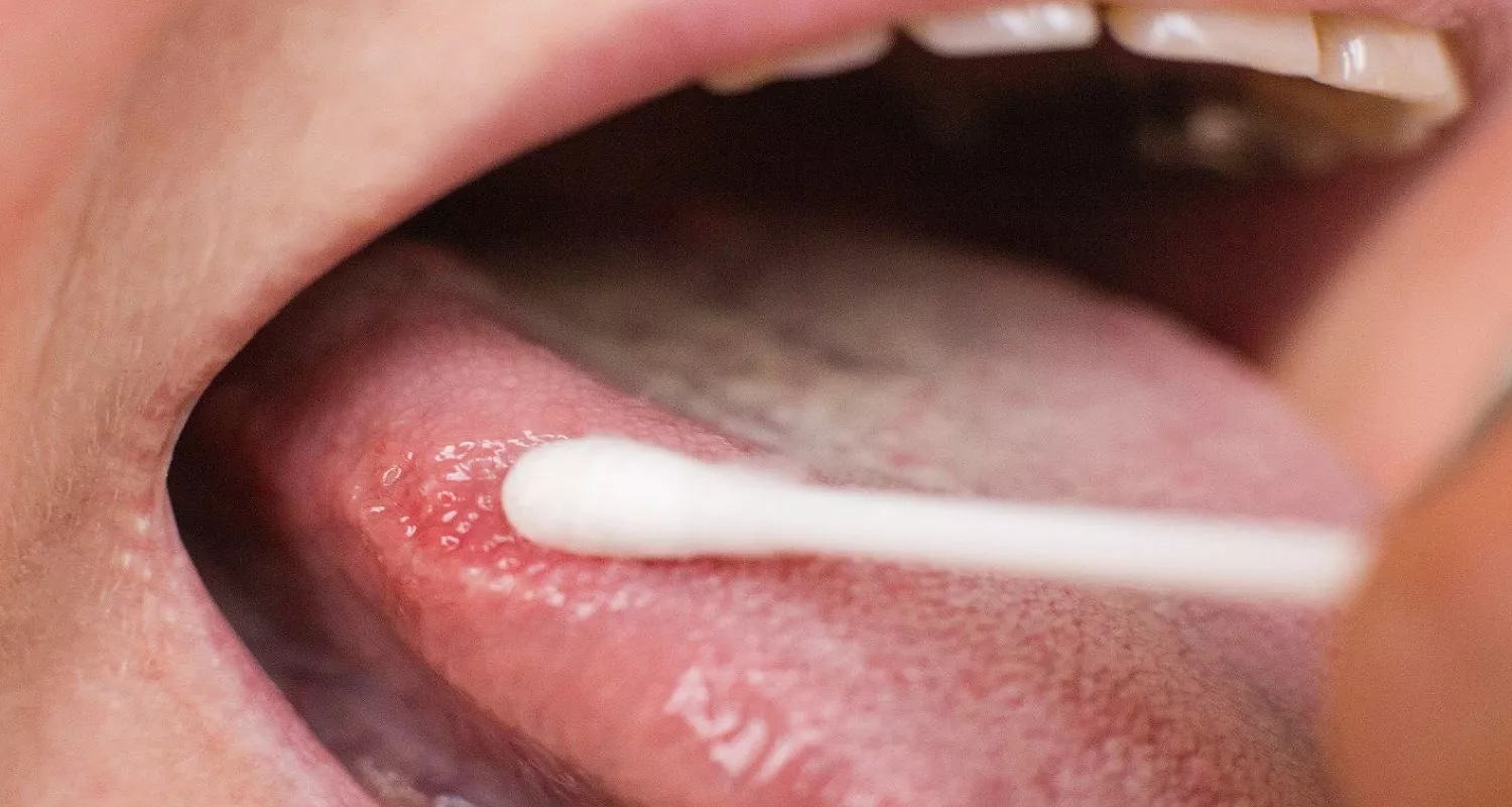
The diagnosis of these lesions is made with a biopsy. The procedure consists of removing a portion of the affected tissue to be observed under a microscope to discern in depth the shape and behavior of these cells. Although a stain with the aforementioned characteristics may constitute a premalignant lesion, it may be due to other diagnoses, in which case the treatment could vary. Among them are:
- Lichen planus
- Oral candidiasis.
- Hemangioma
- Pemphigus
- Leukoedema
- Keratosis
Should I be worried if I have a premalignant lesion?
Although in many cases these lesions, especially leukoplakia, do not turn into cancer, it is important to diagnose them in time to prevent an unfavorable outcome. Generally, in the early stages, the spots can disappear by removing the irritant stimulus. However, adequate follow-up is required in time in case the lesion is not resolved.
Treatment of premalignant lesions
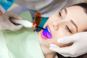
When abnormal lesions are detected in the mouth, the initial treatment consists of removing the stimulus and waiting for the lesion to resolve. If there is no improvement after 2 weeks, the Oral Cancer Foundation recommends taking a biopsy to properly identify the lesion. If it is a premalignant lesion, it can be removed to prevent it from turning into cancer. Some procedures may be indicated, depending upon the severity of the lesion, including:
- Laser surgery
- Cryosurgery
- Cancer treatment: Radiation therapy, chemotherapy, immunotherapy.
Tips to prevent premalignant lesions and
erythroplakia
Many times, premalignant lesions have no known cause. However, the following recommendations may help prevent their appearance:
- Avoid tobacco use.
- Reduce or avoid alcohol consumption.
- Improve oral hygiene.
- Increase consumption of antioxidant foods: fruits, vegetables, and cereals.
- Avoid mouth rinses with alcohol.
- Regularly visit a dentist.
- Check dentures with a dentist every 3 years.
- Replace ill-fitting dentures.


Conclusion
Lesions can occur in the oral mucosa and oropharynx although in many cases they may not represent a significant risk to patients. In others, they may indicate the beginning of a cancerous lesion, especially when it comes to erythroplakia. The Oral Cancer Foundation states that any lesion in the mouth (leukoplakia or erythroplakia) should be considered premalignant until a microscopic examination proves otherwise. Therefore, it is recommended that any anomaly or color change in the oral mucosa by a dentist for a timely diagnosis and proper treatment.
Contact us
If you have any questions about erythroplakia or other topics, you can contact us at Channel Islands Family Dental as well as our page on Facebook. We look forward to your visit and we will make a timely diagnosis. Our dentists in Oxnard, Santa Paula, Ventura, Newbury Park, and Port Hueneme will be able to guide you toward the best treatment to take care of your health and give you back your best smile.
Bibliography
- Diccionario de cáncer del NCI. (s. f.). Instituto Nacional del Cáncer. https://www.cancer.gov/espanol/publicaciones/diccionarios/diccionario-cancer/def/eritroplasia
- Risk Factors – Oral Cancer Foundation | Information and Resources about Oral Head and Neck Cancer. (s. f.). https://oralcancerfoundation.org/understanding/risk-factors/
- Antioxidantes y prevención del cáncer. (Feb 6, 2017). Instituto Nacional del Cáncer. https://www.cancer.gov/espanol/cancer/causas-prevencion/riesgo/dieta/hoja-informativa-antioxidantes
- What Are Leukoplakia and Erythroplakia Lesions? (Dic 19, 2021). WebMD. https://www.webmd.com/oral-health/what-are-leukoplakia-erythroplakia
- ¿En qué consisten los tipos de cáncer de orofaringe y de cavidad oral? (Mar 9, 2018). https://www.cancer.org/es/cancer/cancer-de-orofaringe-y-de-cavidad-oral/acerca/que-es-cancer-de-cavidad-oral.html
- Erythroplakia: Causes, Symptoms &Treatment. (Ene 11, 2023). Cleveland Clinic.https://my.clevelandclinic.org/health/diseases/24595-erythroplakia
- Hennessy B, DDS. Oral proliferations (Last review Feb 2022). Texas A&M University, College of Dentistry. MSD Manual, practitioner version.https://www.msdmanuals.com/professional/dental-disorders/symptoms-of-dental-and-oral-disorders/oral-growths