Our Reviews

Excellent
4.8
I really love this office. It was quick. Clean very friendly. Thank you much.
Response from the owner: Thank you for your kind review
Response from the owner: Thank you
The entire experience was great. The dental assistant or hygienist was very thorough and the dentist was just great. So much better than expected!!
Response from the owner: Hi Matilda thank you so much for your review
Todo el personal fue muy amable. Pero no me gusto porque no me hicieron limpieza de mis dientes en la primera cita sólo radiografía. Lo ciento por mi comentario
Response from the owner: Thank You maleeah for your kind words
Great visit! Everyone was super professional and knowledgeable.
Response from the owner: Thank you !
Response from the owner: Dear Juliette Smith,Thank you for the 5-star rating! We're thrilled you had a positive experience at your trusted Port Hueneme Dentist Channel Islands Family Dental- Port Hueneme. We look forward to serving you again.
Un excelente servicio y muy amables!!
Response from the owner: Muchas gracias por su comentario.
js_loader
Read more reviews

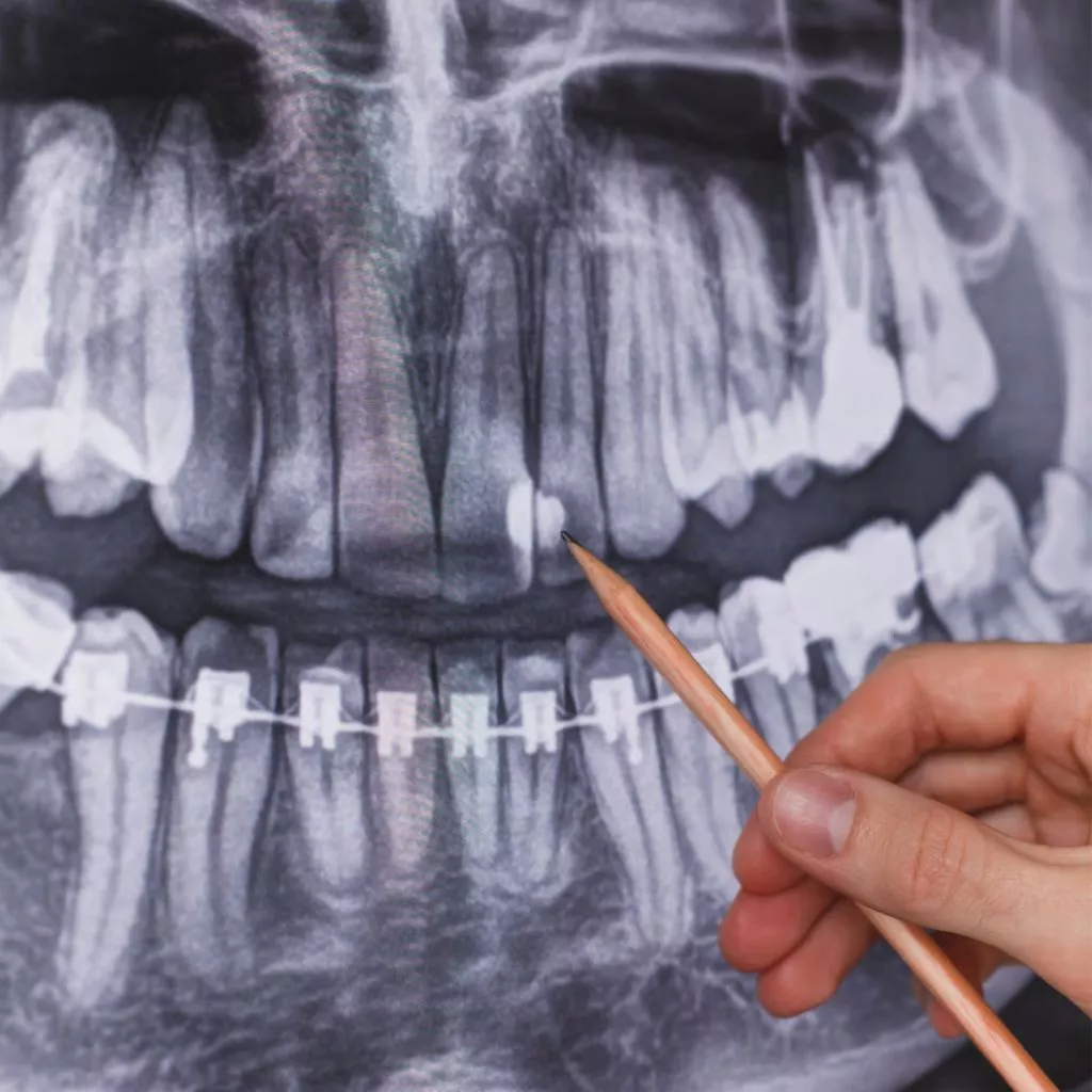
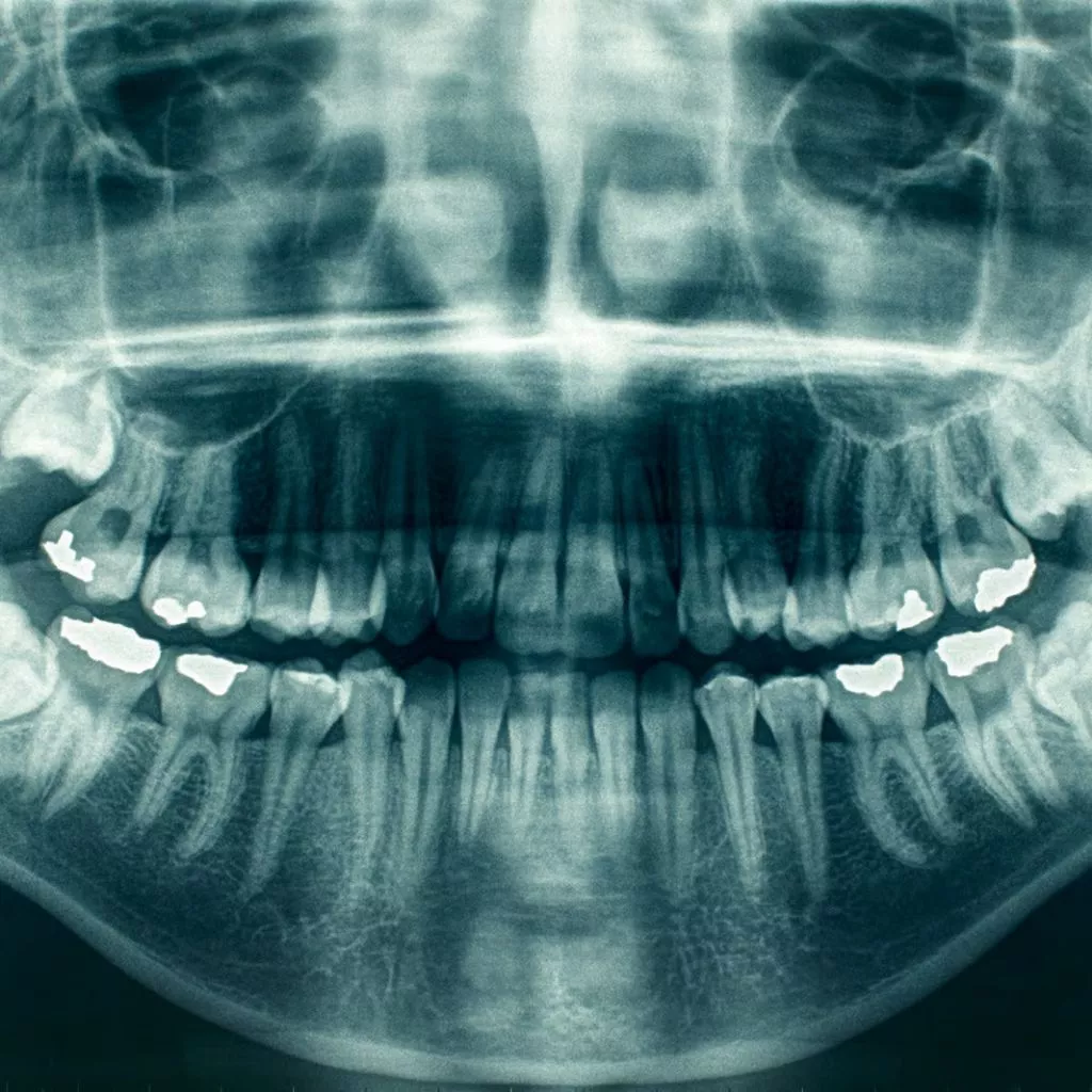
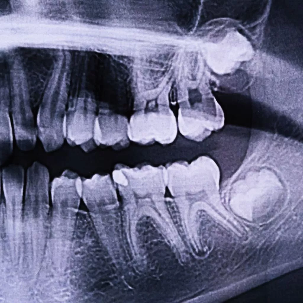
Intraoral Dental Imaging
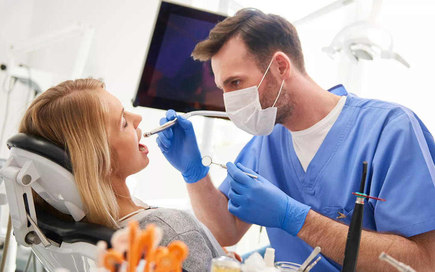
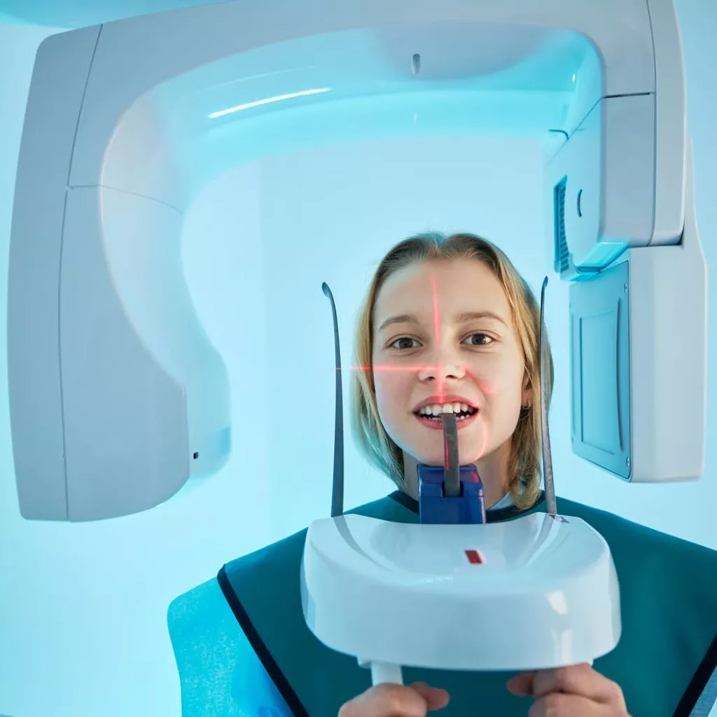
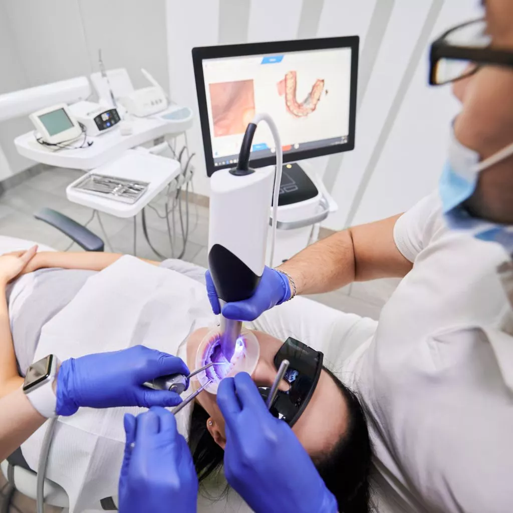
Exploring Intraoral Dental Radiography

Whether it’s for cosmetic enhancement or other motivations, the desire for a flawless, radiant, and whiter smile is on the rise. Technological advancements in dentistry have made achieving this aspiration possible. Presently, there are two popular in-office procedures, both aimed at brightening teeth. While these treatments share similarities in their approach, certain distinctions set them apart.

Information about radiography

INTRAORAL RADIOGRAPHS

Dental images predominantly rely on X-ray technology to provide the essential information needed for making swift and accurate diagnoses. Intraoral radiographs involve the placement of radiographic films inside the mouth, with various types of radiographs available:

  • PERIAPICAL RADIOGRAPHY

Dental radiography, commonly known as this type of radiography, ranks as one of the most commonly sought-after imaging methods in dentistry. It provides a holistic view of the entire tooth, spanning from the root tip to the visible crown in the oral cavity. Through this radiographic image, dental professionals can effectively assess the tooth’s status and the condition of surrounding structures, including the adjacent bone and supporting tissues. Its utility extends to tracking tooth decay advancement, detecting pulp involvement, monitoring infection development, and ruling out fractures or periodontal issues.

  • BITEWING OR INTERPROXIMAL RADIOGRAPHS

Interproximal or bite-wing radiographs, sharing the same dimensions as periapical radiographs, utilize a specialized method to capture intraoral images concentrating on the coronal aspects of the upper and lower dental arches. These radiographs serve as a valuable tool for obtaining information about a wider range of teeth. They are commonly sought to identify dental caries located between teeth, a diagnosis that often eludes clinical examination. Furthermore, they play a crucial role in evaluating the presence and progression of conditions affecting the supporting structures, notably the periodontium.

  • OCCLUSAL RADIOGRAPHY

These radiographs are a tad larger in size in comparison to periapical radiographs. The technique employed to acquire this intraoral image entails the patient biting down on the radiographic plate. Through this radiograph, one can evaluate the positioning of teeth that have not yet erupted within the mouth or those that are situated abnormally.

EXTRAORAL RADIOGRAPHS

intraoral dental imaging

Dental images can be obtained not only through intraoral radiographs but also through extraoral radiographs. The utilization of cone beam computed tomography (CBCT) is now prevalent, offering a wealth of information through three-dimensional (3D) imaging in different planes. A research investigation, detailed in the Journal of Oral and Maxillofacial Surgery and carried out by the academic and professional community associated with the WHO, has confirmed the safety of cone beam tomography, assuring that it complies with rigorous radiation exposure safety standards for patients.

The decision regarding the type of imaging to be requested is guided by multiple factors, with the primary criterion being the nature of the intended observations. Whether the focus is on tooth positioning, periodontal disease identification, fracture or infection diagnosis, or the assessment of treatment outcomes, the choice is dictated by the specific diagnostic task. Importantly, it should be stressed that dental X-ray procedures are classified as safe by the American Dental Association (ADA) owing to their exceedingly low radiation levels, which substantially diminishes the likelihood of potential harm.

INTRAORAL SCANNING

Intraoral imaging of this kind empowers the creation of a 3D digital record of the patient’s oral structure, streamlining digital workflows, and frequently enhancing precision, all while reducing processing time. This modern approach supplants the traditional technique of constructing physical models from the patient’s oral impressions, ultimately enhancing patient comfort.

intraoral dental imaging

Our services

DENTAL EXTRACTIONS

Here, you will find what is a dental extraction, types of dental extractions, how to facilitate recovery, what to do after it, some complications, and a postoperative success

READ MORE

DENTAL IMPLANTS

Here, you will find what is a dental implant, what is the reason of them, how to avoid problems with gums and, some advantages to use them.

Read more

ORTHODONTICS TREATMENT

Get your own perfect smile! Book now with us and get a FREE consultation.

read more
more services

Financing options

Payment plans as low as 99/month (*on approved credit)

Lending Club
Care Credit
Alphaeon Credit

Dental Insurances We Accept

Our Office

Office Hours Monday – Friday: 9:00AM – 6:00PM

book an appointment Call Now (805)-865-7395