Last Updated on: 12th December 2025, 09:41 am
Today, modern dentistry has many advanced tools in dental radiology. This means faster, safer, and more accurate results. Dental X-rays are very important because they help us find problems early and plan better treatments.
In this guide, you will learn how X-rays help our patients in Ventura County, when they are needed, and what safety steps we always follow.
What dental X-rays do we offer in Ventura County?
At Channel Islands Family Dental Office, with locations in Newbury Park, Thousand Oaks, Santa Paula, Port Hueneme, and Camarillo, we take two types of dental X-rays: Periapical X-rays and Panoramic X-rays.
What are periapical or dental X-rays?
Periapical X-rays, also called intraoral X-rays, show a very detailed image of one or more teeth. They help the dentist check the roots, the tooth structure, and the tissues around the tooth.
Our dentists in Ventura County use this type of X-ray often for specific dental problems.
What periapical X-rays are used for
- Finding cavities: Some cavities are small and cannot be seen just by looking. These X-rays help us find them early, so we can treat them before they get worse or painful.
- Checking gum disease: Periapical X-rays help us see if there is bone loss or damage to the tissues that hold your teeth. This is important for diagnosing gingivitis or periodontitis and choosing the right treatment.
- Helping with root canal treatments: During a root canal, these X-rays show us the shape and length of the root canals. They also help us make sure the canals are clean and sealed correctly when we finish the root canal treatment.
- Finding fractures or injuries: If you have trauma or an accident, these X-rays help us look for fractures or teeth that have moved out of place.
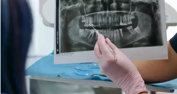
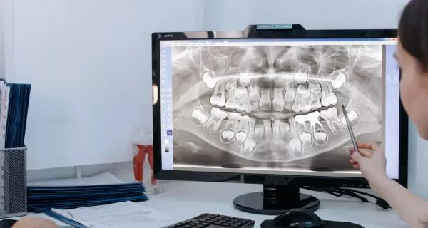
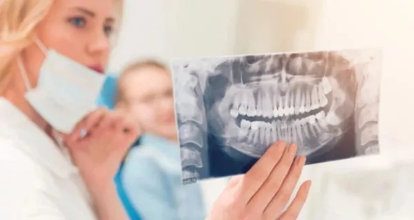
What is a panoramic X-ray?
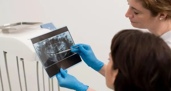
A panoramic X-ray gives a full image of your mouth. It shows all your teeth, your jaw, the joints, and your sinuses. It is not as detailed as a periapical X-ray, but it gives a big-picture view that is very useful for many treatments.
What panoramic X-rays are used for
- Detecting cysts or tumors: This large image helps us see possible cysts, tumors, or other issues in the jaw area.
- Checking dental development in children: A panoramic X-ray shows all the teeth, even the ones that have not come out yet. This helps us see how the teeth are forming and if the position is normal.
- Finding impacted teeth: If a tooth does not come out correctly, it may be impacted. Panoramic X-rays help us locate impacted teeth and plan extractions or orthodontic treatment.
- Evaluating the TMJ (jaw joint): These X-rays help us look at the temporomandibular joint (TMJ) and identify possible problems. In some cases, we may need more detailed tests like a CT scan.
- Planning tooth extractions: A panoramic X-ray shows the position of each tooth and the direction of the roots. This is very important for planning wisdom tooth removal or other extractions safely.
Periapical X-rays vs. panoramic X-rays: what’s the difference?
Both types are useful, but they are used for different reasons.
Detail
- Periapical X-rays: Very detailed images of individual teeth.
- Panoramic X-rays: Show the whole mouth but with less detail.
Uses
- Periapical: Best for cavities, gum disease, root canals, and tooth-specific problems.
- Panoramic: Best for full treatment planning, growth evaluation, and long-term monitoring.
Radiation
- Panoramic X-rays: Use more radiation than one periapical X-ray.
- Both are still considered very safe and low in radiation.
Are dental X-rays safe?
Yes. Dental X-rays are safe for kids, adults, and seniors. At Channel Islands Family Dental Office, we follow strict safety rules. We also use digital X-rays, which use much less radiation than older film X-rays.
We always take the time to explain the process to our patients, especially nervous patients or young children. Many families in Ventura County trust us because we focus on comfort and clear communication during every step.
What safety measures do we use?
- Lead aprons: Patients and dental staff use lead aprons to protect the body and reduce radiation exposure.
- Thyroid collars: We also use thyroid collars to protect the thyroid gland of patients during X-rays.
What equipment do we use?
Our office uses modern digital X-ray technology that produces high-quality images with very low radiation. These systems also help us position the sensor correctly, meaning we rarely need to retake images.
Can I get dental X-rays if I am pregnant?

If you are pregnant or think you may be pregnant, always inform your dentist before taking any X-rays. In most cases, we avoid X-rays unless it is absolutely necessary for an emergency. If the situation is not urgent, we simply postpone them until after pregnancy.
Which type of X-ray do I need?
The type of X-ray you need depends on your dental problem.
At any of our Ventura County offices, Newbury Park, Thousand Oaks, Santa Paula, Port Hueneme, or Camarillo, our dentists will recommend the best option for your situation.
- Periapical X-rays: Best when we need a close, detailed look at one tooth.
- Panoramic X-rays: Best when we need to see the full mouth.
At Channel Islands Family Dental Office, we always choose the X-ray that gives you the safest, most comfortable, and most effective care.